The Local newsletter is your free, daily guide to life in Colorado. For locals, by locals.
GET IT NOW: To see 5280‘s 2012 list of Top Doctors, click here.
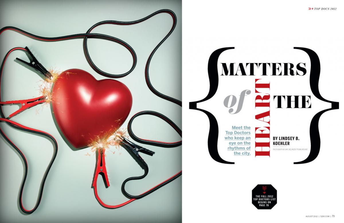
We ascribe much of the human experience to an 11-ounce, cone-shaped muscle that beats rhythmically inside our chests about 100,000 times a day. We are heartsick when tragedy strikes, heartbroken when love goes awry, faint of heart when we lack courage, and heartless when compassion escapes us. Yes, these are turns of phrase—simple constructs of the English language—yet there’s no denying that the one organ we can all feel is just a little more compelling than the ones we can’t. This connection we have with the heart becomes particularly apparent when there’s something wrong with it. It is especially poignant when a heart struggles to beat—and, sadly, struggling hearts are all too common. In fact, 26 percent of Colorado-area deaths—and one in three deaths in America—can be attributed to some form or complication of heart disease. The good news is that the past 25 to 30 years have seen incredible improvements in cardiac care. Today, progress is happening at an exponential rate. And many of Denver’s physicians—including the Top Docs profiled on the following pages—are at the forefront of new treatments, cutting-edge procedures, and promising research into how we can further heal an ailing heart.
Bank On It
Physicians use a lot of terminology with which most of us aren’t familiar. Drug names, physiological processes, human anatomy—hearing a doc speak is often like listening to a foreign language. Pediatric cardiologist and researcher Dr. Shelley Miyamoto knows all the big words, but she’s not above using phraseology that nonmedical types can understand. “We call it ‘freezer diving,’?” she says. “We have this big storage unit where we go to get the heart tissue we need for our lab tests.”
Miyamoto is trying to explain what she does in simple language; however, there’s nothing straightforward about the groundbreaking research she and her team of scientists do. For the past few years, Miyamoto has been the primary investigator for the tissue bank study at Children’s Hospital Colorado. Her research focuses on pediatric heart failure, a condition where the heart doesn’t pump enough blood to oxygenate the body. In kids, the disease usually presents in school-age children, 50 percent of whom will either die or require a heart transplant within five years of diagnosis.
Although the condition is common in adults, it is atypical for a child to have the disease. And because so few children present with heart failure, there has historically been very little research into pediatric-specific medicine. “Almost every treatment we have for heart failure is based on research on adults with heart failure,” Miyamoto says. “What we began to notice is that many drugs that are effective in adults don’t work very well in kids.”
Which brings us back to the freezer. Children’s Hospital Colorado owns the largest pediatric heart tissue bank in the world. To find new drugs—or old drugs that might be used in a new way—to combat pediatric heart failure, Miyamoto needs to examine and work with diseased heart tissue. “When a child receives a heart transplant here at Children’s,” she says, “we always ask if we can take samples from the diseased organ for our tissue bank.” Over the past 17 years, 246 patients have donated pieces of their left and right ventricles, atria, and valves—all of which are housed in a three-foot-by-six-foot unit that keeps hundreds of tiny vials frozen at minus 80 degrees Celsius. That tissue, says Miyamoto, is an invaluable tool in her quest to identify the molecular and mechanical differences between heart failure in kids and heart failure in adults. If things go well in the lab, Miyamoto expects the research to result in clinical trials that could begin helping kids in the next few years. And that’s something we can all understand.
Good Medicine
5280 Why is cardiology compelling to you?
Dr. Chris Lang Cardiovascular medicine has it all in terms of the different dimensions of medicine—preventive care, physiology, biology, surgical procedures, complex imaging.
What are the most common diseases you see?
Atherosclerosis, chronic heart failure, and arrhythmias.
What helps you treat these diseases every day?
We have the benefit of sitting on top of the biggest mound of hard evidence about how to handle problems compared to any other medical specialty. The most clinical trials have been done in cardiovascular medicine because it’s the most common clinical problem. The neat thing is there is always something new being published and there’s always clinical trial data that gives you some real scientific certainty about what you’re advising.
What’s the biggest change you’ve seen in cardiology over the course of your career?
If I look back over the past 20 years, it’s night and day how patients are now faring with coronary artery disease. If you had a heart attack in 1990, your chances of being back in the hospital in five or 10 years with a recurrent event because your atherosclerosis had progressed—despite the fact you were seeing a doctor—were probably a 30 percent chance at five years and a 50 percent chance at 10 years. Today those numbers are dramatically lower. That has to do with prescribing statins (like Lipitor) to treat cholesterol and using antiplatelet agents (like aspirin). Today, I can be confident that after a patient has a heart attack, he can live many years without a recurrence if we are treating him correctly.
Still, more than 500,000 Americans die every year from heart disease. What are the risk factors for atherosclerosis?
The risk factors haven’t changed—age, sex, smoking, diabetes, inactivity, obesity, blood pressure, weight, high cholesterol, sodium intake, and family history.
It’s hard to believe people are still lighting up, isn’t it?
Smoking is still an issue. The most distressing thing as a cardiovascular professional is to see young kids smoking. It drives you out of your mind. You know they’re on a trajectory toward trouble.
The epidemic of diabetes has to be a big issue these days, right?
Looking at national trends in cardiovascular outcomes, the morbidity and mortality have gone down progressively over the last 30 years. But people feel like that curve is now going to take an inflection upward because all that good that’s occurring after a heart attack has the potential to be undone by a wave of new heart disease in individuals who now have diabetes. These people are going to have atherosclerosis that’s related to their diabetes. It would be a shame if cardiovascular disease, which has been recently supplanted by cancer as the primary killer of Americans, regains its ascendancy because of lifestyle issues and the environment we live in.
Plumbing Problems
Dr. James Jaggers likes a challenge. And that’s a good thing, considering the 12 years of postgraduate training he had to complete to do the extremely complicated job he does at Children’s Hospital Colorado. Jaggers is a pediatric cardiothoracic surgeon, which means he takes the hearts of infants into his hands and fixes them every day.
If that doesn’t give you a God complex, nothing will. But Jaggers is humble when he describes how he repairs a life-threatening condition called hypoplastic left heart syndrome (HLHS). “You gotta just do some plumbing really,” he says. “It’s rerouting flows and enlarging tubes. It’s not the most difficult surgery we do, but these HLHS kids are just so sick.”
Babies with HLHS are born with an underdeveloped left side of the heart, which prevents the muscle from pumping enough blood to the body. Without surgery, 90 percent die within one month; 100 percent die within one year. For many years, cardiac-care doctors thought the best way to treat HLHS was an immediate heart transplant. Unfortunately, the average time to receive a pediatric heart transplant is between three and six months. The math just didn’t add up. Instead, surgeons began trying to fix the abnormality surgically, and ultimately figured out that a series of three procedures—in order, the Norwood, the Glenn, and the Fontan procedures—could remedy the problem.
Although the first Norwood procedure was executed in the early 1980s, the procedure was still incurring mortality rates in the 43 percent range well into the late 1990s. Today, many respectable surgical departments are routinely seeing 85 percent survival rates. At Children’s Hospital Colorado, Jaggers and company are hovering around 93 percent, one of the best rates in the country.
A Patient’s Story
Every expectant mother has a love-hate relationship with the 20-week ultrasound appointment. If everything goes well, a mother can find out just how big her baby is getting, the position of the fetus, and whether it’s a boy or a girl. But if everything doesn’t go well, the fun stuff can fade into the background when the technician says something just doesn’t look quite right.
The latter scenario sounds familiar to Teresa Beck, a 28-year-old Loma, Colorado, resident. Pregnant with her first child, Beck was sent to see a specialist when her ultrasound tech told her she wasn’t able to see everything like she wanted to. After visiting with a high-risk pregnancy doctor, Beck had a confirmed diagnosis for her growing baby: hypoplastic left heart syndrome.
Because the HLHS diagnosis was made in utero—about 80 percent are—Beck had to decide whether she wanted to keep the baby. She knew she needed more information. Beck went to Children’s Hospital Colorado. It was there, on December 5, 2011, that she met the crowd of health-care professionals—including Dr. James Jaggers—who could save her child’s life. She decided to have the baby. “There was just no way I could live with the questions,” Beck says. “What if the doctors were wrong? What if it wasn’t as bad as they thought? I decided there must be a reason I was pregnant with a heart baby.”
Because babies with HLHS often need immediate medical intervention after birth, Jaggers wanted Beck to deliver in Denver. So she moved to the Mile High City on January 12, 2012. Curtis Steele was born at 2:05 a.m. on January 23 at University of Colorado Hospital. By 6 a.m. on January 24, one-day-old Curtis was at Children’s Hospital Colorado for emergency surgery to repair complications with some veins surrounding his tiny heart. Eight days later, Jaggers took Curtis to the operating room for the Norwood procedure.
“Curtis came out of the womb looking pretty good,” Jaggers says. “He continued to do really well through the vein surgery and the Norwood.” He did so well, Beck says, the cardiac intensive care unit nurses took to calling Curtis “the rock star baby.”
Of course, even rock stars have ups and downs. Curtis had a few complications, but he was eventually discharged from Children’s in early March 2012. Beck took him home to Loma.
“It’s been nonstop since then,” Beck says. “It’s a full-time job to care for an infant with a heart condition.” She isn’t complaining. Although she will not know the luxury of having a “normal” baby this time around, she says Curtis is doing well and is, of course, a momma’s boy.
Beck took Curtis back to Jaggers for the Glenn procedure in early summer 2012. The Fontan surgery is years away. Curtis’ short-term prognosis is good, though kids with HLHS who undergo this series of procedures do have a much higher risk of eventually developing heart failure as teenagers or adults. But for now, that is a distant worry for Beck. “I don’t know what I would’ve done without Dr. Jaggers,” Beck says. “He’s a miracle worker, and he knows how to make us feel secure in our decisions and our ability to handle all of this.”
Lab Results
Dr. JoAnn Lindenfeld has long been interested in heart failure. She has spent a lengthy and successful career learning about cardiac care, including medicinal intervention, ventricular assist devices, and heart transplants. She helped develop University of Colorado Hospital’s heart transplant department in 1986. She has sat on multiple FDA panels that examine new drugs and novel cardiovascular devices. And she wrote the test (literally) for the advanced heart failure and transplant specialty board certification, one of the newest board certifications in the United States. The doctor still spends much of her days seeing patients in the clinic or in the hospital, but she has recently been carving out time in her sleep-deprived life to venture into medical research. Why? “It’s interesting,” Lindenfeld says. “It allows you to really question things and makes you ask why? and how? and begs you to think deeper about virtually every patient. I think that makes me a better doctor.”
Research, of course, takes many forms, especially at a teaching hospital like University of Colorado Hospital. Not only is Lindenfeld stepping into the lab on occasion, but she also collects patient data to contribute to national databases and other ongoing studies at other institutions. It’s tedious, time-consuming work, but Lindenfeld says there are rewards, even if they are long-term. “Doctors are always frustrated by patients we can’t make better,” she says. “The best days are days when you’re able to be useful and make people better. The most depressing days are when you’ve done everything right and someone doesn’t get better.” Lab time and data collection, she says, are things she can do every day to continue pushing medicine forward. Below, we detail just a small smattering of the current research projects with which Lindenfeld is involved.
{ STUDY HALL }
- Drug Trials Lindenfeld and company are executing early trials of a new drug call Neureglin, which the doctor hopes will help heart failure patients’ hearts squeeze more strongly. Lindenfeld’s work is part of an ongoing national study in which the University of Colorado Hospital is one of 15 participating centers.
- Cardiac Devices University’s heart docs are always researching new types of ventricular assist devices. They are currently studying the effectiveness of the new HeartWare device—which is already approved for bridge to transplant—for long-term use in patients who are not candidates for risky heart transplants.
- Hospital Stays A new study to determine which heart failure patients respond best to diuretics has recently piqued Lindenfeld’s interest. By measuring sodium in patients’ urine after diuretics, she hopes to determine who will respond in the hospital and who will continue to respond when discharged, which could shorten hospital stays for patients.
- Side Effects Funded by the American Heart Association, Lindenfeld is studying hypertension in heart transplant patients, who often develop hypertension due to the effects of immunosuppressive drugs. Lindenfeld is studying the causes of the hypertension as well as the potential effectiveness of a blood pressure drug called Aliskiren.
- The Big C Lindenfeld is involved with a grant submission she hopes will be approved to evaluate cardiac function in women with breast cancer. There are many cancer drugs that can cause heart failure. When heart failure develops, cancer drugs must often be stopped, which means patients do not get the full benefit of the drugs.
Out of Body Experience
A tiny orange-tinted piece of plastic that looks a bit like a small tangerine with legs wobbles on the table. Dr. Max Mitchell picks it up and rolls it slowly and reverently in his hands. He’s familiar with the device’s power. This tiny bubble of plastic, called the Berlin heart, has saved three children in Denver in the past three years. Without it—and without Mitchell’s expertise—those children may not have otherwise had a chance to grow up.
Mitchell has been a pediatric heart surgeon for 13 years, repairing holes in the heart and fixing other congenital defects. In the past couple of years, however, he has been a part of one of the most critical advances in pediatric cardiac care in the United States. When children develop primary heart failure, meaning they have a normal anatomical heart that isn’t working properly, most will require a transplant. But the majority of children with heart failure become very sick waiting on a new heart, which can take several months.
For many years, the adult world of cardiac care has had access to devices that temporarily replace heart function until an organ is found. Until recently, that was not the case in pediatrics. Kids were dying even though the technology to save them existed. “Since the early 2000s, American pediatric heart physicians have been clamoring for a way to bridge kids to transplant,” Mitchell says, explaining that such devices were already available in other countries. “We finally got one approved by the FDA in July 2011.” Mitchell and others at Children’s Hospital Colorado were instrumental in that authorization of the Berlin heart, having participated in the clinical trials.
The Berlin heart is a paracorporeal ventricular device, which means during surgery Mitchell executes a series of steps to connect the device, which rests on the upper abdomen outside the body, to the heart. He attaches a tube to the left side of the heart; that tube comes through the abdominal wall and out to the inflow side of the Berlin heart. The Berlin heart is then connected to a pneumatic pump that vacuum-sucks blood into the ventricle and then pulses positive-pressure air back into the device to push a diaphragm that expels the blood through the outflow cannula, which is connected to the aorta. It’s not a simple process but, as Mitchell explains, having the ability to use this device allows his still-hospital-bound patients to remain in relatively good health until a new heart can be found. “There’s no substitute for the human heart,” Mitchell says, “but this really helps.”
A New Ticker
If you need a new heart, you’ll have to get in line. At any given time, about 10,000 Americans need new hearts. If you’re lucky enough to get one and you live in Colorado, you’ll likely end up at the University of Colorado Hospital, which is the only institution that does heart transplants in our state. Since 1986, the hospital’s heart transplant department has done 417 of the surgeries and boasts a 90.3 percent success rate—slightly higher than the national average. Those stats are not based on luck. They’re the result of a qualified team of cardiac-care docs. Drs. Eugene Wolfel and Joseph Cleveland—an advanced heart failure and transplant cardiologist and a cardiothoracic surgeon, respectively—work with a group of skilled physicians to battle adult heart failure. It may seem like you hear about heart transplants on a regular basis. But the process of treating heart failure and doing heart transplant surgery is a long journey with many detours. Below, Wolfel and Cleveland offer a primer on this medical issue.
{ DID YOU KNOW }
- More than four decades after the first heart transplant took place, the surgery still isn’t all that common. In fact, only about 2,000 of the procedures take place each year in this country.
- Because donor hearts are scarce and there are serious risks associated with any major surgery, the primary goal is to try to treat a patient’s heart failure with medication. For many, meds are successful in controlling their conditions.
- Americans must sign up to be organ donors. Most cardiac-care docs bemoan the fact that in the United States citizens aren’t presumed to be donors like they are in Europe.
- The average wait to receive an adult heart ranges from three to 18 months depending on the status of the patient. That means, the sicker one is, the more likely it is that he or she will receive a new heart.
- A ventricular assist device is a machine that’s partially implanted inside a patient and helps the heart function. About 20 percent of those waiting on a heart transplant get a VAD to help them live until an organ is found.
- VADs are meant to be temporary, but some patients are not good candidates for transplant. VADs can sometimes be used indefinitely. The longest a UCH patient has lived on a VAD is four-and-a-half years after implantation.
- The most common reasons—called contraindications in medical terms—a patient cannot receive a heart transplant are old age and lung disease.
A Patient’s Story
Collin Johnston loves to hike in the Rocky Mountains. When he moved to Colorado in 2005, he quickly became enamored of the trails that wind through the wilds just outside of Boulder. He spent hours on hiking trails—the physical activity at altitude was no trouble for the fit 22-year-old. But after about a year, Johnston began to notice his hikes weren’t quite as effortless. He was short of breath and coughing, and he noticed he’d been gaining weight.
After making a list of his symptoms, Johnston, then 24, surfed common medical sites in an attempt at self-diagnosis. “Every place I looked said my symptoms were all major signs for heart failure,” he says. Johnston was frightened: His mother had died from the disease 13 years earlier.
Johnston’s family doctor diagnosed him with idiopathic dilated cardiomyopathy, which is an enlarged heart with an unknown cause. The disease can have a genetic link; what it doesn’t have is a cure. After an initial regimen of drugs failed to work for Johnston in mid-2006, his doctor in Boulder sent him to University of Colorado Hospital for what he called “alternative treatment.” After seeing the young man, the advanced heart failure docs—a team that consisted of Drs. Eugene Wolfel, JoAnn Lindenfeld, Simon Shakar, Andreas Brieke, and Larry Allen—at University immediately put him on the heart transplant list in November 2006.
For all of 2007 and most of 2008, Johnston managed well on the medications the docs prescribed. He was in and out of the hospital a few times so that doctors could deal with minor complications, but overall things were going well. “I’d go as far as to say that I felt pretty good in early 2008,” he says. “At one point they even deactivated my transplant list status.”
But the good times didn’t last. In December 2008, Johnston deteriorated. He became fatigued, retained fluid, and had trouble sleeping. The doctors upped his meds to no avail. It was time to reconsider a heart transplant, but Johnston was getting sick so quickly that his doctors opted to explore the idea of putting their patient on a ventricular assist device (VAD). “I was really scared about that,” he says, “even knowing Dr. Joe Cleveland was a pro.”
On April 20, 2009, Cleveland implanted the 26-year-old with a HeartMate II, a device that lives almost completely inside a patient’s chest—minus two battery packs and a control unit. The VAD is clunky to wear around every day—Johnston points out, however, that it’s “better than the alternative”—but the unit improved Johnston’s condition immensely. On October 9, 2009, Johnston even walked the Denver Half Marathon, being extra cautious that his battery unit would last the full race.
But VADs aren’t meant to be a permanent fix: Johnston needed a new heart. In August 2009 as well as on December 24, 2009, Johnston got calls for possible hearts—both of which turned out to be false alarms. Finally, his “gift” came on February 3, 2010. It was a perfect match. And after eight hours in the OR, Cleveland had given Johnston another shot at being a weekend warrior. “I can go hiking again,” Johnston says. “I have a lot of prescriptions to take and I have to get my blood drawn all the time, but I think this heart is better than my native heart ever was.”
Screen Saver
If there’s one area of medicine that inspires awe in the operating room, it’s cardiac electrophysiology. Colorful images and wavy lines streak across multiple monitors while a blue-ish three-dimensional rendering of a heart pulses on-screen. On another display, a small video camera reflects internal anatomy in real time. The physician—in this case, Saint Joseph Hospital’s Dr. Laurent Lewkowiez—uses every advanced imaging technology at his disposal to move tiny instruments into the exact right place inside the patient’s heart.
Cardiac electrophysiology is the practice of treating irregularities, called arrhythmias, in the electrical conduction system of the heart. Using intracardiac catheters—small instruments threaded through arteries and veins to the heart—electrophysiologists can place pacemakers or defibrillators, or they can ferret out heart cells that are misfiring and deactivate them using burning or freezing technologies (called ablation). The field has been around for more than 30 years, but recent improvements in high-tech, high-resolution imaging are changing the practice at warp speed. “EP was advancing so rapidly when I came out of training in the late 1990s,” says Lewkowiez, who is a Kaiser Permanente doc, “that I decided to do another fellowship I was not required to do to practice medicine.”
After nine years of post-medical-school training—and now more than a decade of on-the-job experience—Lewkowiez feels more than comfortable treating the variety of maladies that afflict his patients. Things like supraventricular tachycardia (a generally benign but annoyingly fast heartbeat), ventricular tachycardia (a life-threatening rhythm in one of the ventricles), and atrial fibrillation (which increases the risk of stroke and heart failure) are everyday problems that Lewkowiez can solve. “That’s really what’s special about EP,” he says. “You cure people.”
That is, if you’re doing it right. Lewkowiez says that when done well, EP is a minimally invasive procedure with remarkably low complication and mortality rates. But because the field is progressing so quickly, patients need to be proactive when choosing their physicians. “There’s no harm in asking a lot of questions of your electrophysiologist,” he says, “as well as checking that he or she is giving you the right answers.”
Replacement Parts
Modern medicine’s ability to prolong human life has raised the average life expectancy in America to 78.2 years. Better medicines and more refined surgeries are allowing many of us to not only live longer, but also to experience a high quality of life during those “extra” years. The problem is that, just like anything else, the human body has parts that simply wear out after too many years of use.
Ninety-two-year-old Albert Verderaime knows that scenario all too well. Until early April 2012, Verderaime was still working two days a week at the bread bakery his parents opened in 1927 in Aguilar, Colorado. He was baking cakes, Italian bread, loaves of white, and apricot Danishes until he began having chest pains. The aching continued for a couple of weeks before Verderaime decided to visit the Veterans Affairs Medical Center in Raton, New Mexico. “They immediately sent me on a plane to Denver,” Verderaime says.
Without treatment, the aortic stenosis Verderaime was experiencing would likely kill him within the year. The aorta is the main artery that carries blood out of the heart. When blood leaves the organ, it flows through the aortic valve, into the aorta, and then to the rest of the body. With aortic stenosis, the aortic valve does not open fully, causing symptoms like weakness, fatigue, breathlessness, and chest pains. It is fatal within one year in 50 percent of patients with symptoms. What the doctors in New Mexico likely knew before they sent Verderaime north was that he was not a good candidate for open-heart surgery to fix his malfunctioning valve. At the University of Colorado Hospital, however, interventional cardiologists Drs. John Carroll and John Messenger had a better option for Verderaime: transcatheter aortic valve replacement (TAVR).
Approved by the FDA in November 2011, TAVR is a less invasive method of replacing a worn-out valve with a new biologic valve, usually harvested from a cow. Using a catheter system inserted through a three-inch incision in the groin, Carroll and Messenger slowly navigate the valve—which is mounted inside a stent—through the body’s vascular system toward the heart. While the heart is still beating, the doctors can position the stent-mounted valve into place right over the original valve.
“This procedure probably has the largest set of benefits of any new technology in the last decade,” says Carroll. It offers an alternative to people who likely would not survive open heart surgery. It provides an incredible bump in the quality of life for older Americans who might otherwise suffer a miserable final year of their lives. And the possible future applications for this procedure are almost limitless.
Because the procedure is so promising, the University of Colorado Hospital just completed the construction of a $3 million “hybrid” operating room outfitted with a state-of-the-art X-ray system specifically for these surgeries. The OR, which can accommodate the more than 20 health-care professionals it takes to complete this three-hour-long surgery, combines the typical sterile setting of an operating room with the cutting-edge imaging of a cath lab.
For Verderaime, who was Carroll and Messenger’s 10th TAVR patient ever, the procedure was a success. “Mr. Verderaime’s procedure went better than expected,” Messenger says. “He was only in the ICU for 24 hours, and he was discharged just five days after his procedure.” Not only is Verderaime’s chest pain gone, but just weeks after the surgery he is also walking and getting better all the time. And, he says, he hopes to be back at his bakery sooner rather than later.


